Delicate Sense of Touch
Atomic force microscopy supplies researchers and developers extremely high resolution topographical data from a large number of different minerals, polymers, mixtures, composite materials or biological tissue. This technology, developed in the 1980s, enables users to obtain subatomic resolved images of sample surfaces.
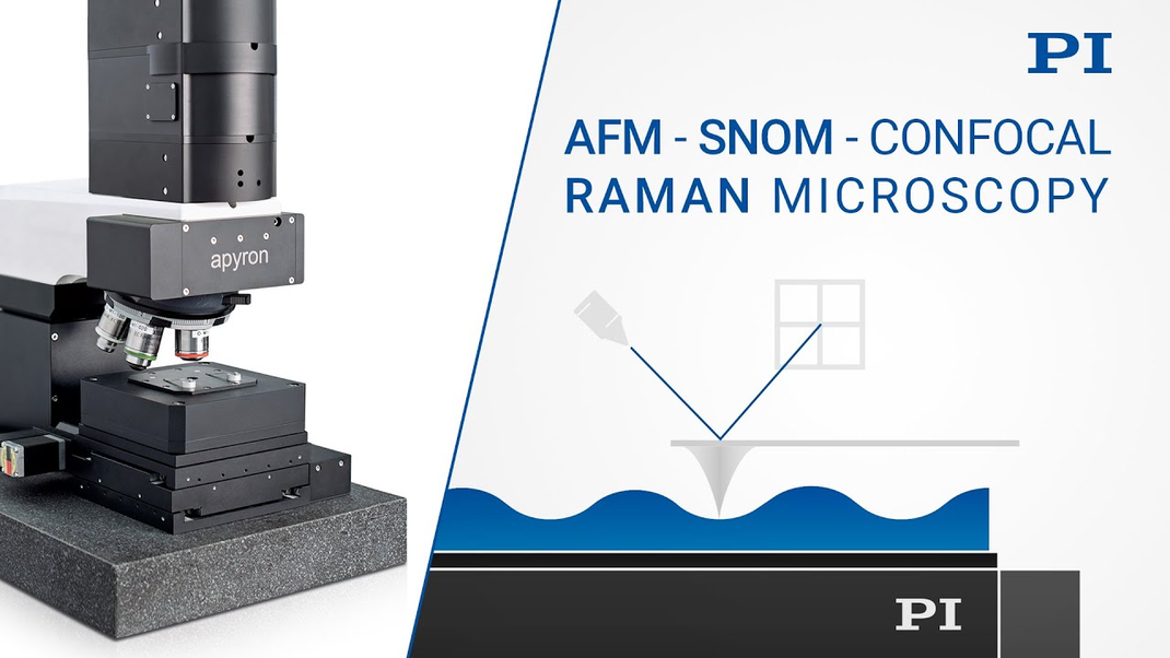
The principle is very simple: A fine needle with a point tapered to the size of just a few atoms is pulled over the sample to be examined. Atomic forces interact between the outermost atom of the needle and the atom of the surface closest to the needle. The effect of the force of the surface on the needle is measured by laser optics.
WITec GmbH, a company based in the German city of Ulm, has developed a special implementation of the principle of obtaining the signal in this way and transferring it to the control of the sample stage. Its Z-position is adjusted so that the same force always acts on the needle. Therefore, the stage position provides the coordinates of the surface topography immediately and the sample stage represents the heart of WITec's atomic force microscopes.
Positioning with the Highest Resolution and Dynamics
The resolution of the sample scanner must be in the subnanometer range because the positioning system provides the spatial resolution. Among other things, the narrow specification is made possible by using capacitive sensors. The mechanical design of the sample table is parallel kinematic. Compared to stacked systems, the trajectories are driven with much more precision and without cumulative errors of individual axes.
At the same time, high dynamics are required because, the faster the topography can be moved in the z direction, the quicker the positioning on the × and y axis. This reduces measuring times and also reduces possible temperature drift which increases with time. The large scanning ranges of up to 200 µm in X and Y are also of great importance.
Dr. Olaf Hollricher, founder and managing director of WITec, confirms: "The combination of a very large scanning range and the high-precision capacitive control means that PI is a unique partner for us". However, the fact that both companies have a similar philosophy is a great advantage for the cooperation that has existed since the foundation of WITec.
Integrating Further Microscopy Modalities
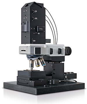
Integrating Further Microscopy Modalities
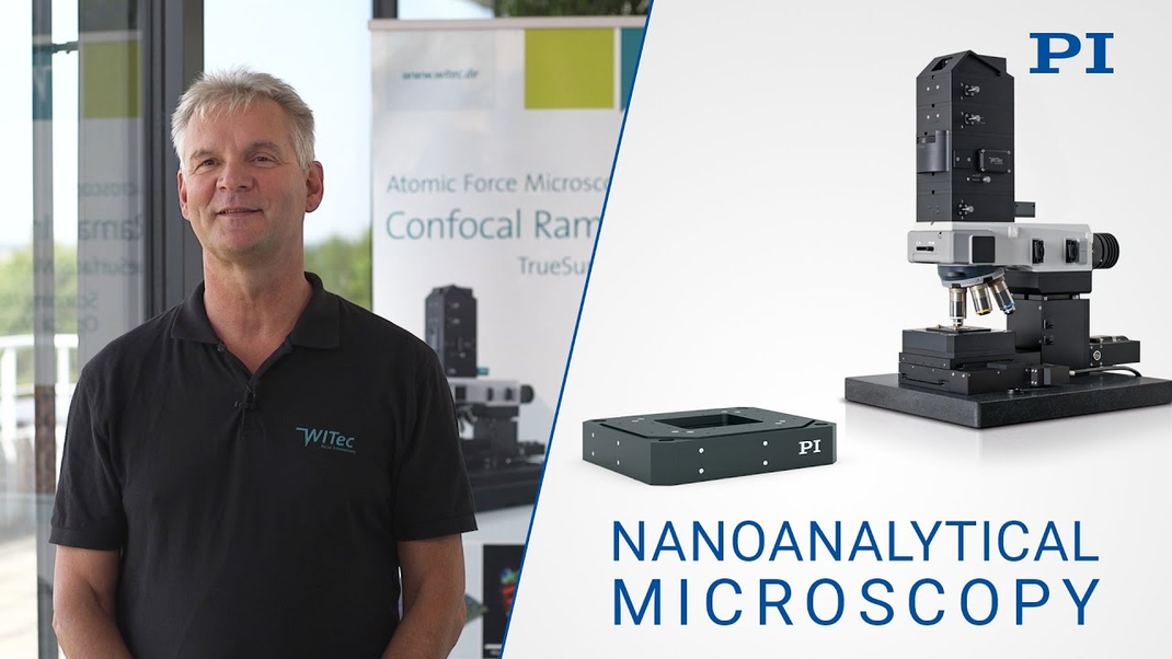
But the WITec system is capable of much more: Two additional microscopy modalities can be combined with atomic force microscopy in the sense of correlative microscopy and displayed together: Raman microscopy and near-field scanning optical microscopy (SNOM). While confocal Raman microscopy identifies the chemical components of the sample, near-field scanning optical microscopy (SNOM) is used for optical imaging with a resolution beyond the diffraction limit. The basis for these extremely high-resolution methods is always the highly precise positioning of the specimen in all three axes of motion.
Key Role of the Scanning Stage
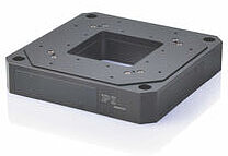
The piezo-based scanning stage plays a key role in specimen positioning. It is designed for travel ranges of 100 or 200 µm along the axes of the scanning plane and 30 µm in direction of the Z axis and allows position resolution better than 2 nm.
In addition, active guiding using capacitive sensors increases path fidelity: The sensors measure any deviations in the perpendicular axis to the direction of motion.
Any undesired crosstalk of the motion can thus be detected and actively compensated in real time. The digital electronics performing the appropriate control also operate at a high cycle rate. This is crucial for the precise assignment of the scanner's position values and the recording camera.
Groundbreaking Results for Countless Scientific Disciplines
Whether forensic analysis, semiconductor technology, material sciences, life sciences or medical technology and this to name but a few, countless disciplines owe groundbreaking insights to atomic force microscopy and its combination with Raman microscopy and SNOM.

